PREPARING FOR TREATMENT
Patient Education
Contact Appointment services at (931) 881-2039
Thank you for choosing Tennessee Heart, and for entrusting us with your cardiology health care needs. We look forward to providing you and your family with comprehensive, quality medical care.
If this is an urgent issue, please call our office. For medical emergencies, or if you are having chest pain, call 9-1-1.

You’re safe with us.
Trusting someone else with your cardiac care is a significant decision. We are here to support you feeling confident in making the best choice for your heart health. The skilled team at Tennessee Heart is dedicated to ensuring that you receive the best possible care available.
Cardiac Echo
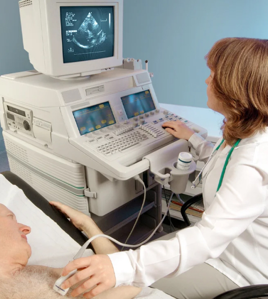
Echocardiography, also called an echo test or heart ultrasound, is a test that takes moving pictures of the heart with sound waves. You do not have to stay in the hospital. It’s not surgery and does not hurt.
Your doctor may use an echo test to look at your heart’s structure and check how well your heart is working.
This test may be needed if:
• You have a heart murmur.
• You’ve had a heart attack.
• You have unexplained chest pains.
• You’ve had rheumatic fever.
• You have a congenital heart defect.
Trained sonographers typically conduct echo tests in a variety of settings, including doctors’ offices, emergency rooms, operating rooms, hospital clinics, and hospital rooms. During the test, you’ll lie on a bed on your left side or back while the sonographer applies a special jelly to a probe and moves it over your chest area. The probe uses ultra-high-frequency sound waves to create images of your heart and valves, without the use of X-rays. You can watch the images of your heart’s movements on a video screen, and the sonographer can also take a videotape or photograph of the pictures.
The test is painless, has no side effects, and typically takes around one hour to complete.
In some cases, a closer view of your heart is required to obtain clearer images, which may necessitate a special test known as transesophageal echocardiography (TEE). During this test, a cardiologist will pass a tube with a probe on the end of it down your throat and into your esophagus while you swallow. The probe will then use sound waves to create images of your heart, as described above. After the test is complete, the cardiologist will gently remove the probe, and you may feel the need to cough.
- The size and shape of your heart
- How well your heart is working overall
- If a wall or section of heart muscle is weak and not working correctly
- If you have problems with your heart’s valves
- If you have a blood clot

Transesophageal Echocardiogram (TEE)
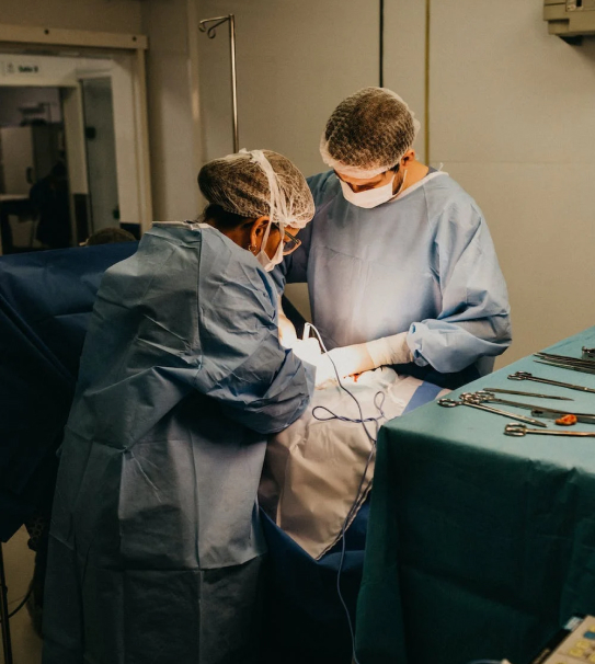
Your doctor may recommend a transesophageal echocardiogram for several reasons. The most common is to provide additional information during heart surgery or cardiac catheterization.
A TEE is performed when the standard echocardiogram isn’t clear enough to make the suspected diagnosis. It’s also performed in patients who are having heart surgery to give the surgeon and anesthesia team more information to guide treatment after surgery and confirm that the surgical procedure has been successful or if additional repair is needed prior to leaving the operating room. The risk of a TEE is minimal and your cardiologist will discuss with you the reasons you need a TEE as well as standard echocardiography.
There are two main advantages of this type of echocardiogram. First, it allows your cardiologist to get a much better look at some of the heart structures, including the wall between the two top chambers of the heart (atrial septum) and heart valves. This is because the esophagus and stomach are very close to the heart and may allow the cardiologist to obtain more detailed pictures of the heart compared to routine echocardiograms performed from the chest wall.
Secondly, it allows the cardiologist to obtain images of the heart during heart procedures (surgery and cardiac catheterization) without having the echo camera get in the way of the procedure. This procedure may cause discomfort and gagging if done without sedation.
The procedure usually takes 20 to 40 minutes, but the total time with sedation is usually 60 to 90 minutes. The procedure is very safe. Your doctor will go over the risks of the procedure with you and give you an opportunity to ask questions prior to the test. Almost everyone can return to normal activity within 24 hours

Treadmill stress test
A stress test, sometimes called a treadmill test or exercise test, helps your doctor find out how well your heart handles its workload. As your body works harder during the test, it requires more fuel and your heart has to pump more blood. This test can show if there’s a lack of blood supply through the arteries that go to the heart.
It also helps your doctor know the kind and level of physical activity that is right for you.
Doctors use exercise stress tests to find out a number of things:
- If you have an irregular heartbeat.
- If your symptoms (such as chest pain or difficulty breathing) are related to your heart.
- How hard you should exercise when you are joining a cardiac rehabilitation program or starting an exercise program.
- If treatments you have received for heart disease are working.
- If you need other tests (such as a coronary angiogram) to detect narrowed arteries.
Tell your doctor about any medicines (including over-the-counter, herbs and vitamins) you take.
- He or she may ask you not to take them before the test. Don’t stop taking them unless the doctor says to.
- You may be asked not to eat, drink or smoke for two to four hours before the test. You may drink water.
- Wear comfortable, loose-fitting clothing and walking shoes with rubber soles. Shorts or sweatpants and jogging or tennis shoes are good choices.
- You’re hooked up to equipment to monitor your heart.
- You walk slowly in place on the treadmill.
- It tilts so you feel like you’re going up a small hill.
- It changes speeds to make you walk faster.
- You may be asked to breathe into a tube for a couple of minutes.
- You can stop the test at any time if you need to.
- After slowing down for a few minutes, you’ll sit or lie down and your heart and blood pressure will be checked.
- Your heart rate
- Your breathing
- Your blood pressure
- Your electrocardiogram (ECG or EKG)
- How tired you feel What equipment is used? The electrocardiography machine will record your heartbeat and heart waves in an electrocardiogram (ECG).
- Wires, or electrodes, will be hooked up to your chest and arms or shoulders. The wires are connected to the ECG machine.
- Near the end, you may breathe into a mouthpiece that will measure the air you breathe out.
There’s very little risk — no more than if you walked fast or jogged up a big hill. Medical professionals are on hand in case anything unusual happens during the test.

